Biomolecules — Practice Quiz
Tamil Nadu Board · Class 12 · Chemistry
Try a 4-question quiz on Biomolecules for Tamil Nadu Board Class 12 Chemistry: tap an answer to check it and see why.
Interactive on Super Tutor
Studying Biomolecules? Get the full interactive chapter.
Quizzes, flashcards, AI doubt-solver and a step-by-step study plan — built for practice quiz and more.
Free trial, no card needed.
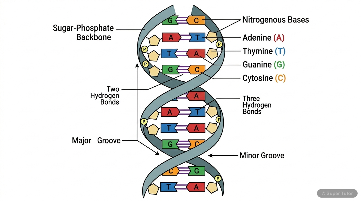
An illustration from Super Tutor's Biomolecules chapter — alongside flashcards, concept maps and practice questions.
Quick Quiz: Biomolecules
0/4Tap an answer to check it instantly. No sign-up needed for these 4.
Glucose on oxidation with bromine water gives gluconic acid, but on oxidation with concentrated nitric acid gives glucaric acid (saccharic acid). What structural information does this provide about glucose?
The phenomenon of mutarotation in glucose involves which of the following sequences of events?
Sucrose is called an invert sugar after hydrolysis. Which of the following correctly explains this term and the chemistry behind it?
Which statement correctly distinguishes the glycosidic bonds and reducing sugar properties of sucrose, lactose, and maltose?
Sample Questions
In amylopectin, branching occurs every 24–30 glucose units through α-1,6 glycosidic bonds, while in glycogen, branching occurs every 8–14 glucose units. What is the biological significance of more frequent branching in glycogen?
Show answer
More frequent branching in glycogen creates more free non-reducing ends, allowing faster enzymatic hydrolysis and quicker glucose release during high metabolic demand.
Step 1: Both amylopectin and glycogen are branched polysaccharides made of α-D-glucose units with α-1,4 bonds in chains and α-1,6 bonds at branch points. Step 2: Each branch end is a non-reducing end where enzymes (glycogen phosphorylase) can act. More branches = more non-reducing ends = more simultaneous sites for enzymatic action. Step 3: Glycogen (branching every 8–14 units) has MANY more branch ends than amylopectin (every 24–30 units). This means glycogen can be degraded MUCH faster, releasing glucose rapidly when energy is needed (e.g., during exercise or fight-or-flight response). Step
An amino acid at a pH BELOW its isoelectric point will exist predominantly in which ionic form, and what is its behaviour in an electric field?
Show answer
It exists as a cation (positively charged, –NH₃⁺ and –COOH groups) and migrates toward the cathode (negative electrode) in an electric field.
Step 1: The isoelectric point (pI) is the specific pH at which an amino acid has ZERO net charge (zwitterion form: –NH₃⁺ and –COO⁻). Step 2: At pH BELOW pI → the solution is more acidic → excess H⁺ ions are present → the –COO⁻ group gets protonated back to –COOH → the amino acid carries a net POSITIVE charge (cationic form: –NH₃⁺ and –COOH). Step 3: A positively charged ion (cation) migrates toward the CATHODE (negative electrode) in electrophoresis. Step 4: At pH ABOVE pI → solution is more basic → the –NH₃⁺ group loses a proton to become –NH₂ → net NEGATIVE charge (anionic form) → migrates t
Which statement correctly describes the hydrogen bonding pattern in an α-helix secondary structure of proteins?
Show answer
The carbonyl oxygen (C=O) of the nth residue forms a hydrogen bond with the amino hydrogen (N–H) of the (n+4)th residue, giving 3.6 residues per turn with a 5.4 Å pitch.
Step 1: In the α-helix, the polypeptide backbone winds in a right-handed helical spiral. The key feature is the specific hydrogen bonding pattern in the MAIN CHAIN (backbone), NOT in side chains. Step 2: The C=O of residue n forms a hydrogen bond with the N–H of residue n+4. This means every fourth residue along the chain is hydrogen bonded to each other. Step 3: This creates a very regular, stable helical structure with exactly 3.6 amino acid residues per complete turn. Each turn spans 5.4 Å (0.54 nm) along the helix axis. Step 4: The R groups (side chains) protrude OUTWARD from the helix and
Haemoglobin has a quaternary structure while myoglobin has only tertiary structure. Which of the following statements BEST explains the structural and functional difference between these two proteins?
Show answer
Haemoglobin has four polypeptide subunits (quaternary structure) allowing cooperative oxygen binding, while myoglobin is a single polypeptide chain (tertiary structure) that stores oxygen in muscles.
Step 1: QUATERNARY STRUCTURE is defined as the arrangement of multiple polypeptide chains (subunits) into a single functional unit. A protein MUST have more than one polypeptide chain to have quaternary structure. Step 2: HAEMOGLOBIN consists of FOUR polypeptide chains (2 α-chains + 2 β-chains), each with a heme group containing iron. The four subunits interact with each other → QUATERNARY STRUCTURE. This allows cooperative binding — as O₂ binds to one subunit, it changes the shape of others making them bind O₂ more easily (sigmoidal binding curve). Step 3: MYOGLOBIN is a SINGLE polypeptide ch
+41 more questions on Biomolecules (Tamil Nadu Board Class 12 Chemistry)
Practise AllFrequently Asked Questions
What are the important topics in Biomolecules for Tamil Nadu Board Class 12 Chemistry?
How many practice questions are there for Biomolecules?
Sources & Official References
Content is aligned to the official syllabus. Refer to the board website for the latest curriculum.
More resources for Biomolecules
For serious students
Get the full Biomolecules chapter — start free.
Quizzes, flashcards, an AI doubt solver and a study plan for Tamil Nadu Board Class 12 Chemistry. Free to start, no card needed.