Human Reproduction — NCERT Solutions
Madhya Pradesh Board · Class 12 · Biology
NCERT Solutions for Human Reproduction, Madhya Pradesh Board Class 12 Biology: 21 textbook questions solved step by step.
Interactive on Super Tutor
Studying Human Reproduction? Get the full interactive chapter.
Quizzes, flashcards, AI doubt-solver and a step-by-step study plan — built for NCERT solutions and more.
Free trial, no card needed.
Learn better with visuals Super Tutor pairs illustrations like this with notes and quizzes for Human Reproduction.
The first 11 solutions are open to read. The other 10 are free with a Super Tutor account.
EXERCISES — Human Reproduction
1Fill in the blanks:
(a) Humans reproduce __________ (asexually/sexually)
(b) Humans are __________ (oviparous, viviparous, ovoviviparous)
(c) Fertilisation is __________ in humans (external/internal)
(d) Male and female gametes are __________ (diploid/haploid)
(e) Zygote is __________ (diploid/haploid)
(f) The process of release of ovum from a mature follicle is called ___________
(g) Ovulation is induced by a hormone called ___________
(h) The fusion of male and female gametes is called ___________
(i) Fertilisation takes place in ___________
(j) Zygote divides to form ___________ which is implanted in uterus.
(k) The structure which provides vascular connection between foetus and uterus is called ___________Show solution
(a) Humans reproduce sexually.
(b) Humans are viviparous (they give birth to young ones directly, without laying eggs).
(c) Fertilisation is internal in humans (it occurs inside the female reproductive tract).
(d) Male and female gametes are haploid (they contain half the chromosome number, ).
(e) Zygote is diploid (formed by fusion of two haploid gametes, ).
(f) The process of release of ovum from a mature follicle is called ovulation.
(g) Ovulation is induced by a hormone called Luteinising Hormone (LH) (the LH surge triggers ovulation).
(h) The fusion of male and female gametes is called fertilisation.
(i) Fertilisation takes place in the ampulla of the fallopian tube (oviduct).
(j) Zygote divides to form blastocyst which is implanted in the uterus.
(k) The structure which provides vascular connection between foetus and uterus is called placenta.
2Draw a labelled diagram of male reproductive system.Show solution
Diagram of Male Reproductive System
Although a hand-drawn diagram is required in the board exam, the key structures to be labelled are described below:
Key structures and their positions:
- Testes — paired oval organs located in the scrotal sac; site of spermatogenesis and androgen production.
- Epididymis — coiled tube on the posterior surface of each testis; site of sperm maturation and storage.
- Vas deferens (Ductus deferens) — muscular tube that carries sperms from epididymis upward into the pelvic cavity.
- Seminal vesicles — paired glands that secrete fructose-rich fluid forming part of semen.
- Prostate gland — single gland surrounding the urethra; secretes slightly alkaline fluid.
- Bulbourethral glands (Cowper's glands) — paired glands that secrete lubricating fluid.
- Urethra — common passage for urine and semen.
- Penis — male external genitalia; contains erectile tissue.
- Scrotum — pouch of skin housing the testes, maintains temperature ~2–3°C below body temperature.
- Ejaculatory duct — formed by union of vas deferens and duct of seminal vesicle.
Note to student: Draw a neat diagram showing the above structures in correct anatomical positions and draw arrows with labels for each part. The urethra should be shown passing through the prostate and penis.
3Draw a labelled diagram of female reproductive system.Show solution
Diagram of Female Reproductive System
Key structures to be labelled in the board exam diagram:
- Ovaries — paired organs located in the pelvic cavity; produce ova and ovarian hormones (estrogen, progesterone).
- Fallopian tubes (Oviducts) — paired tubes connecting ovaries to uterus; each has:
- Infundibulum — funnel-shaped opening with finger-like fimbriae that collect the ovum.
- Ampulla — wider region; site of fertilisation.
- Isthmus — narrow region joining the uterus.
- Uterus (Womb) — pear-shaped muscular organ; site of implantation and foetal development.
- Cervix — narrow lower part of uterus opening into vagina.
- Vagina — fibromuscular tube connecting cervix to the exterior; birth canal.
- External genitalia — mons pubis, labia majora, labia minora, clitoris, hymen.
- Mammary glands — shown separately; produce milk after parturition.
Note to student: Draw a neat mid-sagittal section view showing ovaries connected to the uterus via fallopian tubes. Label all parts with clear leader lines.
4Write two major functions each of testis and ovary.Show solution
Functions of Testis:
- Spermatogenesis: The seminiferous tubules of the testis produce male gametes (spermatozoa) by the process of spermatogenesis.
- Androgen secretion: The Leydig cells (interstitial cells) present in the testis synthesise and secrete male sex hormones called androgens (primarily testosterone), which regulate the development of secondary sexual characters and accessory reproductive structures.
Functions of Ovary:
- Oogenesis: The ovary produces female gametes (ova/eggs) by the process of oogenesis.
- Hormone secretion: The ovary secretes female sex hormones — estrogens (by developing follicles) and progesterone (by corpus luteum) — which regulate the menstrual cycle, development of secondary sexual characters, and maintenance of pregnancy.
5Describe the structure of a seminiferous tubule.Show solution
Structure of a Seminiferous Tubule:
Given: Each testis has about 250 testicular lobules, and each lobule contains 1–3 highly coiled seminiferous tubules.
Structure:
- Each seminiferous tubule is a highly coiled, long tubule lined by a germinal epithelium.
- The inner wall (lumen side) of the tubule is lined by two types of cells:
- Spermatogonia (male germ cells): These are the primary male germ cells. They are large, rounded cells lying near the basement membrane. They undergo mitotic and meiotic divisions to ultimately produce spermatozoa (spermatogenesis).
- Sertoli cells (nurse cells): These are large, columnar cells extending from the basement membrane to the lumen. They provide nutrition to the developing germ cells. They also secrete inhibin, which provides feedback control of spermatogenesis, and androgen-binding protein (ABP).
- The basement membrane surrounds the tubule from outside.
- Outside the seminiferous tubules, in the interstitial spaces, lie the Leydig cells (interstitial cells), which synthesise and secrete androgens (testosterone).
- Also present in the interstitium are blood vessels, lymph vessels, and connective tissue.
Summary: The seminiferous tubule is the functional unit of the testis where spermatogenesis occurs, supported by Sertoli cells, while Leydig cells in the surrounding interstitium produce androgens.
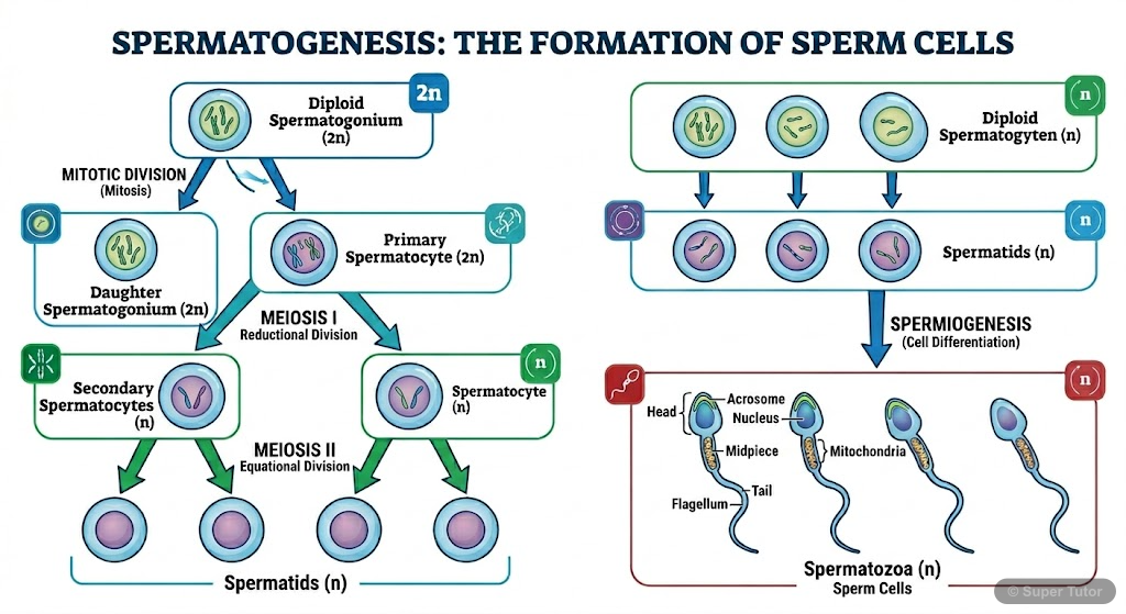
6What is spermatogenesis? Briefly describe the process of spermatogenesis.Show solution
Definition:
The process of formation of spermatozoa (male gametes) from the spermatogonial cells (primary germ cells) in the seminiferous tubules of the testis is called spermatogenesis.
Process of Spermatogenesis:
Step 1 — Multiplication phase:
- The undifferentiated germ cells called spermatogonia () are present on the inner wall of seminiferous tubules.
- They undergo repeated mitotic divisions to increase in number.
Step 2 — Growth phase:
- Some spermatogonia (called primary spermatocytes, ) grow in size by accumulating nutrients.
Step 3 — Meiotic phase:
- Each primary spermatocyte () undergoes Meiosis I (first meiotic division) to form two secondary spermatocytes (), each with half the chromosome number.
- Each secondary spermatocyte undergoes Meiosis II (second meiotic division) to form two spermatids ().
- Thus, one primary spermatocyte gives rise to 4 spermatids.
Step 4 — Spermiogenesis:
- The spermatids are transformed into mature, motile spermatozoa (sperms) by a process called spermiogenesis.
- This involves differentiation: formation of head, neck, middle piece, and tail.
Step 5 — Spermiation:
- The mature sperms are released from the Sertoli cells into the lumen of the seminiferous tubule — this is called spermiation.
Hormonal regulation: Spermatogenesis is initiated at puberty due to increased secretion of GnRH from the hypothalamus, which stimulates the anterior pituitary to secrete FSH and LH. FSH stimulates Sertoli cells to facilitate spermatogenesis; LH stimulates Leydig cells to secrete testosterone, which is essential for spermatogenesis.
7Name the hormones involved in regulation of spermatogenesis.Show solution
The following hormones regulate spermatogenesis:
- Gonadotropin-Releasing Hormone (GnRH): Secreted by the hypothalamus at puberty; stimulates the anterior pituitary to release gonadotropins (FSH and LH).
- Follicle Stimulating Hormone (FSH): Secreted by the anterior pituitary; acts on Sertoli cells and stimulates them to secrete factors that help in spermatogenesis.
- Luteinising Hormone (LH) / ICSH (Interstitial Cell Stimulating Hormone): Secreted by the anterior pituitary; acts on Leydig cells and stimulates them to secrete testosterone.
- Testosterone (Androgens): Secreted by Leydig cells of the testis; essential for the initiation and maintenance of spermatogenesis.
- Inhibin: Secreted by Sertoli cells; provides negative feedback to the anterior pituitary to inhibit FSH secretion, thereby regulating the rate of spermatogenesis.
Summary: GnRH → FSH + LH → Sertoli cells (spermatogenesis) + Leydig cells (testosterone) → spermatogenesis.
8Define spermiogenesis and spermiation.Show solution
Spermiogenesis:
The process of transformation of non-motile, rounded spermatids into mature, motile spermatozoa (sperms) is called spermiogenesis.
During spermiogenesis, the following changes occur in the spermatid:
- The nucleus condenses and elongates to form the head.
- The Golgi apparatus forms the acrosome (cap-like structure over the nucleus).
- The centrioles form the flagellum (tail).
- Mitochondria aggregate around the proximal part of the flagellum to form the middle piece.
- Most of the cytoplasm is shed.
Spermiation:
The process by which mature spermatozoa are released from the Sertoli cells into the lumen of the seminiferous tubule is called spermiation.
After spermiation, the sperms are transported through the male accessory ducts (rete testis → vasa efferentia → epididymis → vas deferens) to the exterior during ejaculation.
9Draw a labelled diagram of sperm.Show solution
Labelled Diagram of a Human Sperm:
A human sperm is approximately 60 µm long and consists of four parts:
1. Head:
- Contains the elongated, haploid nucleus with densely packed chromatin.
- Covered anteriorly by a cap-like structure called the acrosome, which is derived from the Golgi apparatus.
- The acrosome contains hydrolytic enzymes (e.g., hyaluronidase, acrosin) that help in penetration of the egg during fertilisation.
2. Neck:
- Short region connecting the head to the middle piece.
- Contains the proximal centriole (helps in formation of the first cleavage spindle after fertilisation).
3. Middle piece:
- Contains numerous mitochondria arranged in a spiral (mitochondrial sheath) around the axial filament.
- Mitochondria provide energy (ATP) for sperm motility.
4. Tail (Flagellum):
- Long, whip-like structure responsible for the motility of the sperm.
- Contains the axoneme (9+2 arrangement of microtubules).
Note to student: Draw an elongated sperm showing the four regions clearly. Label: Acrosome, Nucleus (Head), Neck, Mitochondrial sheath (Middle piece), Axial filament, Tail/Flagellum.
10What are the major components of seminal plasma?Show solution
Seminal plasma is the fluid component of semen (excluding spermatozoa). It is secreted by the male accessory glands.
Major components of seminal plasma:
- Fructose — secreted mainly by seminal vesicles; provides energy (nutrition) to the spermatozoa.
- Citric acid — secreted by the prostate gland.
- Calcium and certain enzymes — secreted by the prostate gland; help in coagulation and liquefaction of semen.
- Prostaglandins — secreted by seminal vesicles; help in sperm transport by stimulating uterine contractions.
- Mucus / lubricating fluid — secreted by bulbourethral glands (Cowper's glands); lubricates the urethra and neutralises the acidic urine residue.
- Bicarbonate buffers — maintain the slightly alkaline pH of seminal plasma (~7.4), which protects sperms from the acidic environment of the vagina.
Function: Seminal plasma provides nutrition, protection, and a suitable medium for the transport and survival of spermatozoa.
11What are the major functions of male accessory ducts and glands?Show solution
Major Functions of Male Accessory Ducts:
| Duct | Function |
|---|---|
| Rete testis | Collects sperms from seminiferous tubules and passes them to vasa efferentia. |
| Vasa efferentia | Transport sperms from rete testis to epididymis. |
| Epididymis | Site of maturation and storage of spermatozoa; sperms gain motility and fertilising capacity here. |
| Vas deferens | Transports sperms from epididymis to the ejaculatory duct; also stores sperms. |
| Ejaculatory duct | Carries sperms and secretions of seminal vesicles into the urethra. |
| Urethra | Common passage for semen and urine; carries semen to the exterior during ejaculation. |
Major Functions of Male Accessory Glands:
- Seminal vesicles: Secrete a fructose-rich, alkaline fluid that provides energy to sperms and constitutes about 60% of semen volume.
- Prostate gland: Secretes a slightly alkaline, milky fluid containing citric acid, calcium, and enzymes; helps in coagulation and subsequent liquefaction of semen; neutralises vaginal acidity.
- Bulbourethral glands (Cowper's glands): Secrete a mucus-like lubricating fluid that lubricates the urethra and neutralises any residual acidic urine before ejaculation.
Overall function: The accessory ducts transport sperms, while the accessory glands produce seminal plasma that nourishes, protects, and facilitates the transport of spermatozoa.
Free with a Super Tutor account
Free with a Super Tutor account
Free with a Super Tutor account
(a) Corpus luteum
(b) Endometrium
(c) Acrosome
(d) Sperm tail
(e) Fimbriae
Free with a Super Tutor account
(a) Androgens are produced by Sertoli cells. (True/False)
(b) Spermatozoa get nutrition from Sertoli cells. (True/False)
(c) Leydig cells are found in ovary. (True/False)
(d) Leydig cells synthesise androgens. (True/False)
(e) Oogenesis takes place in corpus luteum. (True/False)
(f) Menstrual cycle ceases during pregnancy. (True/False)
(g) Presence or absence of hymen is not a reliable indicator of virginity or sexual experience. (True/False)
Free with a Super Tutor account
Free with a Super Tutor account
Free with a Super Tutor account
Free with a Super Tutor account
Free with a Super Tutor account
Free with a Super Tutor account
10 more solved questions in Human Reproduction
They are free with a Super Tutor account, along with practice quizzes and flashcards for this chapter. Free to start, no card needed.
Frequently Asked Questions
What are the important topics in Human Reproduction for Madhya Pradesh Board Class 12 Biology?
Are these NCERT Solutions for Human Reproduction free?
How should I revise Human Reproduction for the Madhya Pradesh Board Class 12 board exam?
Sources & Official References
Content is aligned to the official syllabus. Refer to the board website for the latest curriculum.
More resources for Human Reproduction
Practice Quiz
Test yourself with a quick quiz
Important Questions
Exam-style questions with answers
Revision Notes
Key points for last-minute revision
Chapter Summary
Understand the chapter at a glance
Concept Maps
See how topics connect
Study Plan
Step-by-step plan for this chapter
Flashcards
Quick-fire cards for active recall
Syllabus
What topics to cover
For serious students
Get the full Human Reproduction chapter — start free.
Quizzes, flashcards, an AI doubt solver and a study plan for Madhya Pradesh Board Class 12 Biology. Free to start, no card needed.